 Promethean
The Brief
A brain-imaging study published in Molecular Psychiatry has mapped for the first time how ketamine produces its rapid antidepressant effects in people with treatment-resistant depression, identifying region-specific changes in glutamate receptor density that correlate directly with symptom improvement. The research, led by Yokohama City University, used a novel PET tracer across 83 participants to visualise molecular changes previously observable only in animal models.
The Report
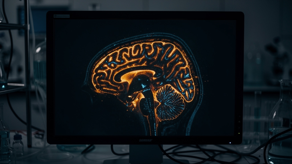
Researchers at Yokohama City University have produced the first direct evidence in living humans of the molecular mechanism behind ketamine’s fast-acting antidepressant effects, using positron emission tomography to track how the drug reshapes glutamate receptor distribution across specific brain regions in patients with treatment-resistant depression.
The study, published March 5 in Molecular Psychiatry, integrated data from three registered clinical trials conducted in Japan, scanning 34 patients with treatment-resistant depression and 49 healthy controls. Patients received intravenous ketamine or placebo over a two-week period, with PET scans performed before and after treatment using a tracer called [11C]K-2 — developed by the same team — that enables visualisation of cell-surface AMPA receptors in the living brain.
The results revealed that ketamine does not produce uniform brain-wide changes. Instead, it drives region-specific modulation of AMPA receptor density: increasing receptor levels in cortical regions responsible for complex cognition, while decreasing them in the habenula, a structure involved in processing disappointment and aversive experience. The pattern of these molecular shifts correlated directly with reductions in depressive symptoms. Greater receptor changes corresponded with better clinical outcomes.
“Using a novel PET tracer, [11C]K-2, we were able to visualize how ketamine alters AMPAR distribution across specific brain regions and how these changes correlate with improvements in depressive symptoms,” said Professor Takuya Takahashi, who led the research. The changes, he noted, “partially rescued” the receptor abnormalities observed in depressed patients relative to healthy controls.
The findings arrive seven years after the FDA’s 2019 approval of esketamine nasal spray — the first glutamate-targeting treatment for depression — and amid growing clinical use of ketamine for patients who do not respond to conventional antidepressants. Roughly 30 percent of people diagnosed with major depressive disorder meet the criteria for treatment resistance, representing an estimated 2.8 million adults in the United States alone. Standard antidepressants typically require four to six weeks to take effect. Ketamine can produce measurable relief within hours.
The study’s limitations include a small sample size, a two-week follow-up window, and a participant cohort drawn entirely from Japan. The lead researcher holds a patent application related to the PET tracer used and is founder of AMPAMETRY, Inc., which holds exclusive licensing rights to it.
The researchers proposed that AMPA receptor PET imaging could eventually serve as a biomarker for predicting individual patient response to ketamine — a step toward what the field increasingly describes as precision psychiatry.
The Angle
The significant finding here is not that ketamine works — that has been observed clinically for years. It is that the mechanism is region-specific and measurable. Cortical regions gain receptor density. The habenula loses it. The brain is not being uniformly stimulated. It is being selectively reconstructed, with different regions moving in opposite directions according to a pattern that maps onto symptom improvement.
That distinction matters because it changes what the next generation of treatments can aim at. A drug that produces diffuse neurochemical change is a blunt instrument — effective, but with side effects baked into its imprecision. A drug designed against a specific receptor profile in a specific region is something closer to repair. The distance between those two approaches is the distance between managing a condition and understanding it well enough to reverse it.
The broader pattern is worth noting. For decades, psychiatric pharmacology operated largely in the dark — drugs were approved because they worked in trials, not because anyone could watch the mechanism operate in a living brain. That era is closing. The ability to image molecular changes at this resolution, in real time, in the organ that produces consciousness, is not an incremental improvement in psychiatric research. It is the beginning of psychiatry becoming an engineering discipline — one that can see what it is fixing.
The first imaging tools precise enough to watch a drug rebuild synaptic architecture in a living human brain are not the end of a research programme. They are the specification sheet for everything that comes after.